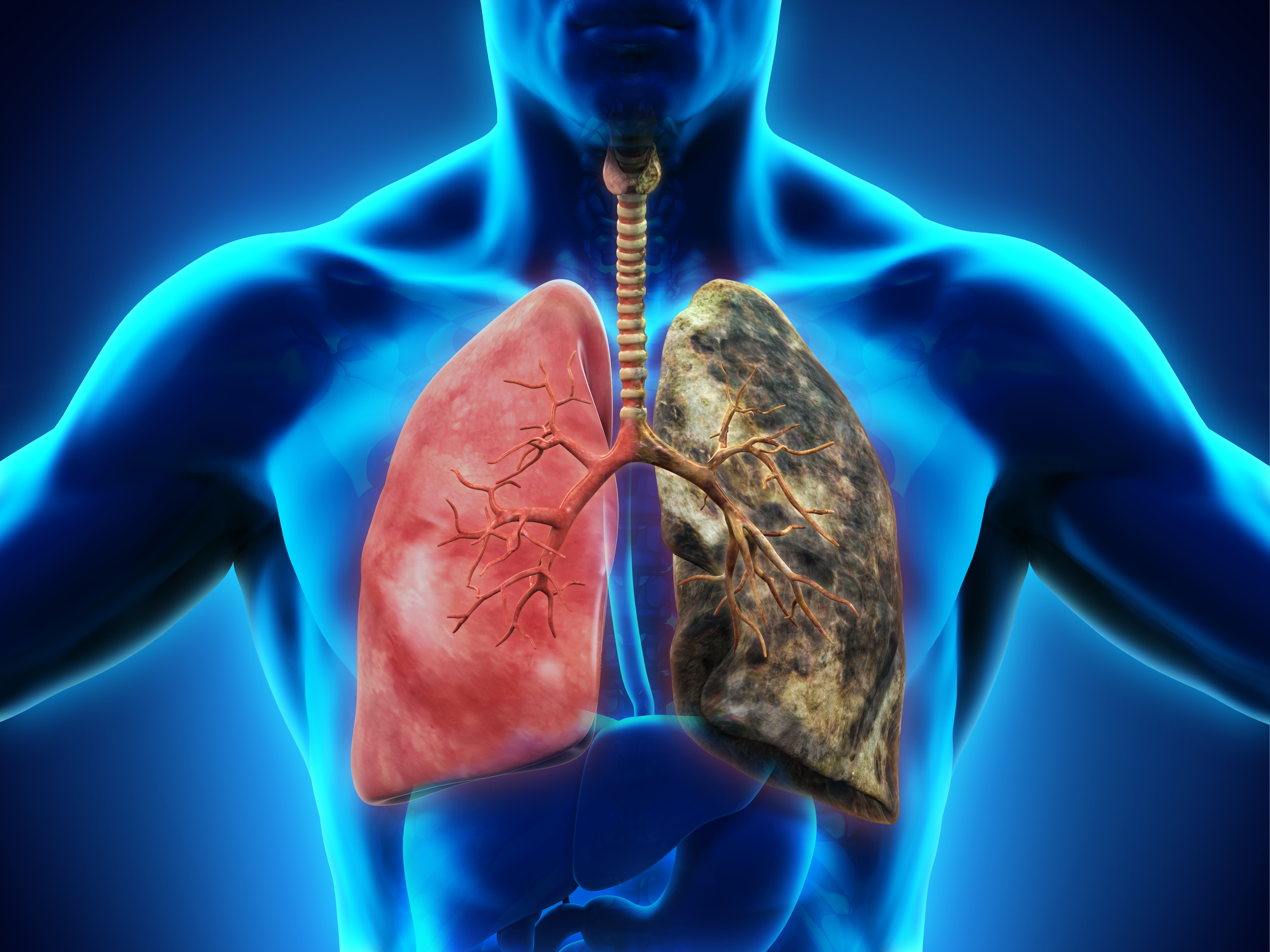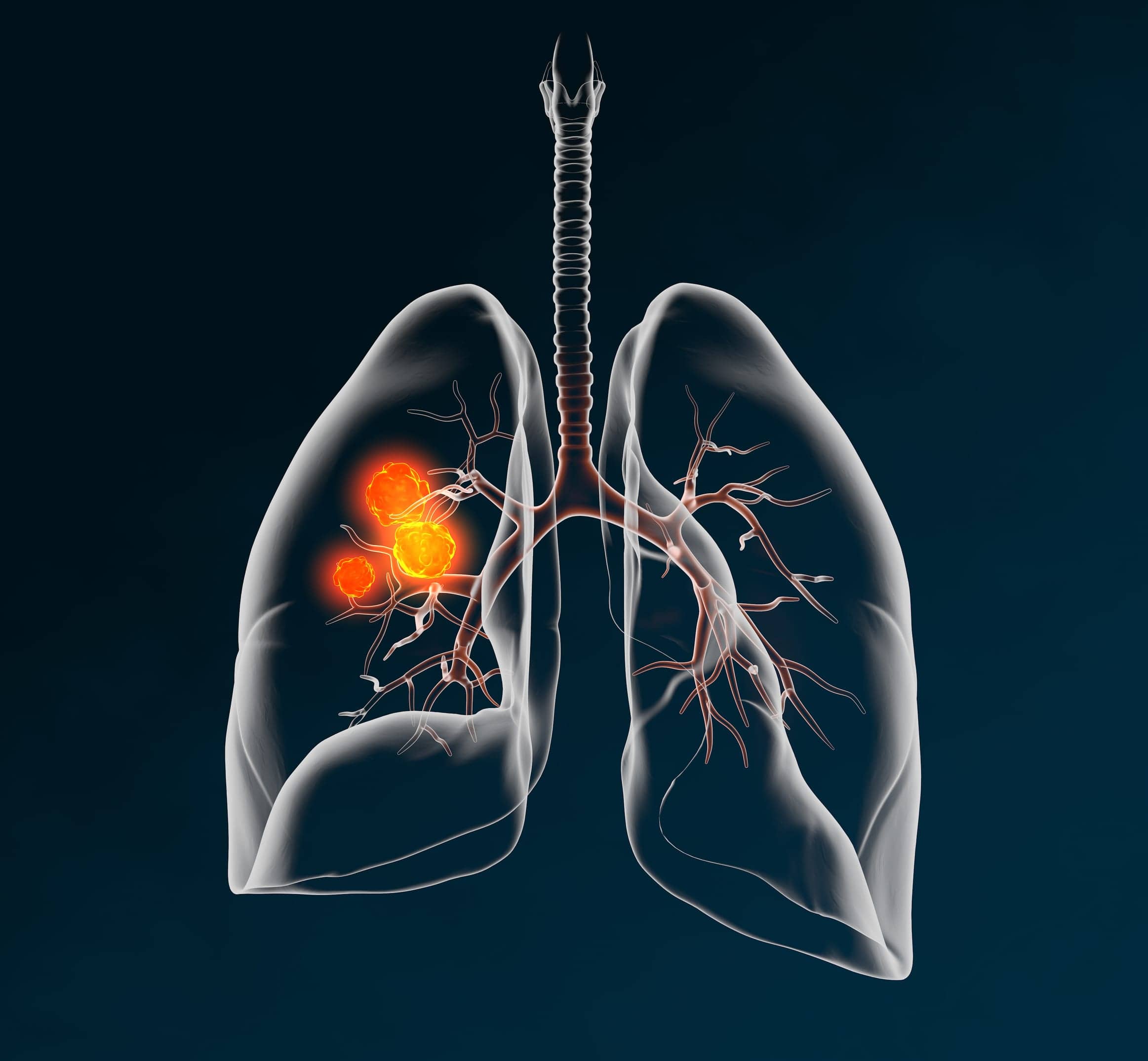
Lung cancer pictures images

cancer of the right lung. Notice the white mass in the middle portion of the right lung (seen on the left side of the picture). Review Date 8/3/ A CT scan uses multiple X-ray images to create a detailed view of the lungs. It is more reliable than an X-ray for showing lung tumors. It can also show the. Imaging tests to look for lung cancer. Imaging tests use x-rays, magnetic fields, sound waves, or radioactive substances to create pictures of the inside of. The big picture. Lung cancer has been the most common cancer in the world for several decades, accounting for. 1 in 5 of all cancer deaths1. The data are organized as “collections”; typically patients' imaging related by a common disease (e.g. lung cancer), image modality or type (MRI, CT, digital.
NCI Visuals Online contains images from the collections of the National Cancer Institute's Office of Communications and Public Liaison, including general biomedical and science-related images, cancer-specific scientific and patient care-related images, and portraits of directors and staff of the National Cancer Institute. Mar 16, · In the current study, we show that ART1 is expressed on the surface of human lung cancer cells and that its expression is associated with reduced lung tumor infiltration of P2X7R + CD8 T cells. In preclinical models of lung cancer and melanoma, ART1 expression on tumor cells promoted escape from CD8 T cell–mediated tumor control. Mar 12, · High Risk of Lung Cancer. Over age Nodule is larger than 3 cm in diameter. Patient smokes or is a former smoker. Exposure to occupational toxins such as asbestos or radon. First- or second-degree relative with lung cancer. Presence of lung cancer symptoms such as persistent cough or shortness of breath.
While traditional X-rays do help detect lung cancer, they offer less detailed pictures from one angle as compared with more advanced imaging technology. Cancerous lesions can often be distinguished from benign lesions on chest CT scans. Your doctor cannot diagnose cancer with only an image from a CT scan or an X. Lung Cancer Europe (LuCE) is the voice of people affected by lung cancer in Lung Cancer Europe, profile picture. Log In.
Nov 19, · Benign. Mediastinal lymph nodes are located along the lining of the lung and, like all lymph nodes, can become enlarged during www.viborgsky.ru can sometimes be read as a spot on an X-ray. Benign tumors can also develop in the lungs, the most common of which are tissue malformations called hamartomas. Other types of benign tumors include fibromas, bronchial . Nov 05, · COPD Causes. Smoking and secondhand smoke plays a significant role in causing COPD. About 85% to 90% of all COPD deaths are related to smoking. The other causes are related to environmental irritants (pollution), and a rare few are genetically passed through family members (for example, people with Alpha-1 antitrypsin deficiency [AAT] are more likely . Jun 21, · Lung cancer is one of the most common malignant cancers worldwide and bears the 2 mL of serum‐free medium was added into each well and the pictures were taken by phase‐contrast microscope (Nikon Microphot‐FX) at 0, 24, and 48 h. (Beyotime). Finally, images were taken by a fluorescence microscope (Olympus). THP‐1.
In most cases of early lung cancer, there are no symptoms, and the cancer may be discovered on imaging tests performed for unrelated reasons. cancer of the right lung. Notice the white mass in the middle portion of the right lung (seen on the left side of the picture). Review Date 8/3/
Nov 05, · COPD Causes. Smoking and secondhand smoke plays a significant role in causing COPD. About 85% to 90% of all COPD deaths are related to smoking. The other causes are related to environmental irritants (pollution), and a rare few are genetically passed through family members (for example, people with Alpha-1 antitrypsin deficiency [AAT] are more likely . Non-small-cell lung cancer is the most common type of lung cancer. X-rays use low doses of radiation to make images of structures inside your body. . Jun 21, · Lung cancer is one of the most common malignant cancers worldwide and bears the 2 mL of serum‐free medium was added into each well and the pictures were taken by phase‐contrast microscope (Nikon Microphot‐FX) at 0, 24, and 48 h. (Beyotime). Finally, images were taken by a fluorescence microscope (Olympus). THP‐1.
In most cases of early lung cancer, there are no symptoms, and the cancer may be discovered on imaging tests performed for unrelated reasons. Cancer types · Bladder cancer · Breast cancer · Cancer of the uterus · Cervical cancer · Colorectal cancer · Head and neck cancer · Lung cancer. A CT scan uses multiple X-ray images to create a detailed view of the lungs. It is more reliable than an X-ray for showing lung tumors. It can also show the. The big picture. Lung cancer has been the most common cancer in the world for several decades, accounting for. 1 in 5 of all cancer deaths1.
Mar 12, · High Risk of Lung Cancer. Over age Nodule is larger than 3 cm in diameter. Patient smokes or is a former smoker. Exposure to occupational toxins such as asbestos or radon. First- or second-degree relative with lung cancer. Presence of lung cancer symptoms such as persistent cough or shortness of breath. Nov 05, · COPD Causes. Smoking and secondhand smoke plays a significant role in causing COPD. About 85% to 90% of all COPD deaths are related to smoking. The other causes are related to environmental irritants (pollution), and a rare few are genetically passed through family members (for example, people with Alpha-1 antitrypsin deficiency [AAT] are more likely . Feb 07, · Adenocarcinoma is the most common form of lung cancer. It's generally found in smokers. However, it is the most common type of lung cancer in nonsmokers. It is also the most common form of lung cancer in women and people younger than As with other forms of lung cancer, your risk of adenocarcinoma increases if you. Smoke.
While traditional X-rays do help detect lung cancer, they offer less detailed pictures from one angle as compared with more advanced imaging technology. Browse 3, lung cancer stock photos and images available, or search for lungs or cancer to find more great stock photos and pictures. Advanced or metastatic lung cancer may be diagnosed through these procedures: Magnetic resonance imaging (MRI) scans create detailed images of inside the body. This slide show features images of CT-guided biopsies, transthoracic ultrasounds, PET/CT scans, and x-rays revealing non–small-cell lung cancer (NSCLC). See a picture of and learn about lung cancer, a type of malignancy, in the eMedicineHealth Image Collection Gallery.
Apr 06, · Colon cancer is a disease in which malignant (cancer) cells form in the tissues of the colon. The colon is part of the body’s digestive www.viborgsky.ru digestive system removes and processes nutrients (vitamins, minerals, carbohydrates, fats, proteins, and water) from foods and helps pass waste material out of the www.viborgsky.ru digestive system is made up of the esophagus, .: Lung cancer pictures images
| Lung cancer pictures images | 687 |
| Lung cancer pictures images | |
| Amazon jigsaw puzzles |
VIDEO
Chest xray - Pneumonia, tumor or something else
In my opinion, it is actual, I will take part in discussion. I know, that together we can come to a right answer.
I apologise, but, in my opinion, you are not right. I can defend the position. Write to me in PM.
It to it will not pass for nothing.
What excellent topic
You commit an error. I can prove it. Write to me in PM, we will communicate.